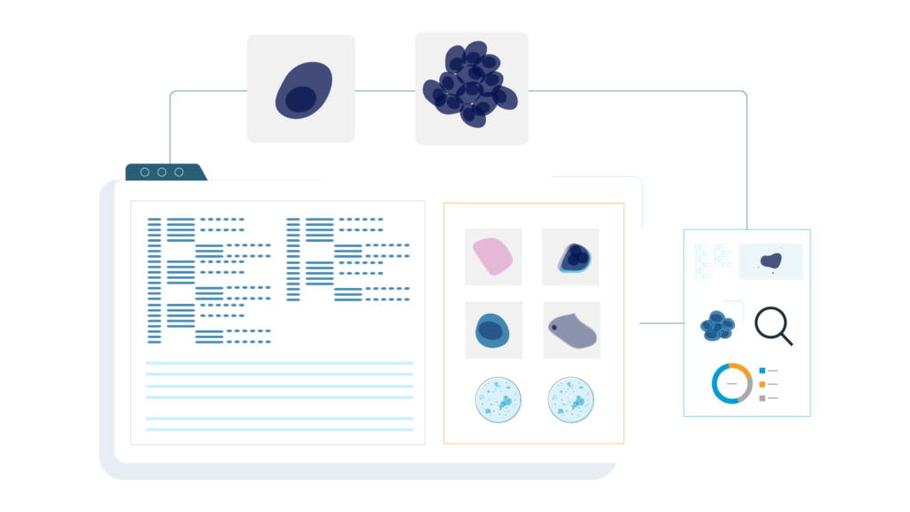
VisioCyt® uses deep learning and computer vision algorithms to analyze and provide a diagnosis on digitized cytology slides.
VisioCyt® facilitates the early detection of cancer indicators, providing pathologists with a valuable tool to refine their diagnoses.
Originating from French research
Protected by five patents
Advantages
AI is revolutionizing cytological analysis
Automated and reproducible analysis
Thanks to its algorithms, VisioCyt® performs morphological analysis of each cell, reducing interpretation biases.
Improved diagnostic accuracy
By using advanced algorithms, VisioCyt® increases the sensitivity of detecting cellular abnormalities compared to traditional cytology.
Scalable solution
VisioCyt® continuously improves its performance through the accumulation of analyzed data.
How it works
The VisioCyt® algorithms
The VisioCyt® algorithms automatically analyze cytology images to detect morphological features associated with cellular abnormalities.
They combine image processing and machine learning techniques to provide a cancer diagnosis.
Quality control
Validation of slide preparation according to the correct protocol.
Detection & classification
Identification of biological elements, classification, and selection of objects of interest.
Additional quality control
Verification of a sufficient number of objects of interest among all detected biological elements.
Analysis of objects of interest
Characterization and classification of detected cells based on more than 10 atypia criteria.
Slide classification
Diagnosis based on cell types and their characteristics. A linear classifier generates a score between 0 and 1 for the slide.
WORKFLOW
VisioCyt® in pathology laboratories
Schéma Anatomopathologie
Urine Sample
Urine sample sent to the laboratory for analysis.
Cytology Slide
VisioCyt® is compatible with standard staining techniques used in pathology laboratories.
Slide Scanner
High-resolution digitization of the cytology slide.
Machine Learning & Deep Learning
Automated analysis by artificial intelligence remotely.
Pathologists & Cytotechnicians
VisioCyt® transmits the analysis results to the pathologist.
SOLUTIONS
Our solutions for cancer detection
Based on VisioCyt® technology, several products have been developed for different types of cancer, providing healthcare professionals with reliable, high-performance tools specifically tailored to each pathology.